How to perform a femoral artery exposure
Source
To access the full video and additional content on this subject, log in or subscribe
Summary
Focus: Safe surgical exposure and vascular control of the proximal femoral arteries.
Key elements: Identification of the common femoral artery, superficial femoral artery, deep femoral artery, femoral vein, femoral nerve branches, inguinal ligament, and bifurcation level.
Indications and Clinical Context
General
Medical experts: Ville Vänni (trauma surgeon)
Name of procedure: Femoral artery exposure
Goal of Operation
To expose and gain vascular control of common femoral, superficial femoral, and deep femoral arteries for treatment.
Problem
Hemorrhage or limb ischemia due to vascular injury or occlusive disease.
Diagnosis
Trauma/injury
Unspecified injury of femoral artery S75.01
Ischemia
Embolism and thrombosis of arteries of the lower extremities I74.3
Unspecified atherosclerosis of native arteries of extremities I70.20
Short Pathophysiological Description
Femoral artery injuries occur through direct trauma (iatrogenic puncture, penetrating injury, blunt force) or iatrogenic access complications that damage the vessel wall. Arterial puncture too proximally above the femoral head prevents effective compression against bone, while through-and-through punctures might create posterior wall bleeding that is inaccessible for direct compression. The common femoral artery's short length, approximately 5 cm, and its position as the sole blood supply continuation to the lower extremity means that uncontrolled hemorrhage or thrombosis threatens limb viability.
Key Anatomical Structures
Common femoral artery
Superficial femoral artery, or femoral artery
Deep femoral artery
Femoral circumflex arteries
Femoral vein
Deep femoral vein
Femoral circumflex veins
Great saphenous vein
Femoral nerve and its branches
Inguinal ligament
Anterior superior iliac spine (ASIS)
Pubic tubercle
Step-by-Step Technique
Patient Positioning, Anesthesia and Preparation
The patient is positioned supine with the groin exposed.
Anesthesia choice depends on the procedure, being typically general or spinal anesthesia, or local anesthesia for purely endovascular procedures.
Sterile preparation and draping depend on the indication. For vascular injuries, the lower abdomen is included in the sterile field to allow proximal extension for external iliac artery access if needed.
Approach Goal and Strategy
The goal is to access and gain control of the proximal femoral arteries: the common femoral artery, superficial femoral artery, and deep femoral artery.
Using the longitudinal femoral approach, this is accomplished by incising through skin and subcutaneous tissue, dividing the fascia layer, identifying the common femoral artery, and exposing its bifurcation into the superficial and deep femoral arteries.
Important structures to avoid injuring include the femoral nerve branches laterally to the artery and the femoral vein medially.
Log in or subscribe to access full content and see all the images.
Landmarks and Incision Site
The external iliac artery becomes the common femoral artery at the inguinal ligament level, then bifurcates a few centimeters distally into the superficial femoral artery (SFA), the primary vessel to the lower extremity, and deep femoral artery, or profunda femoris, which supplies thigh collateral circulation.
The inguinal ligament extends from the anterior superior iliac spine (ASIS) to the pubic tubercle. The palpation finding of the ligament on the skin, or the inguinal crease, is typically felt slightly more distal to the actual ligament.
The skin incision is positioned at the inguinal ligament level, approximately midway between the ASIS and pubic tubercle. The artery lies just medial to the midpoint of the inguinal ligament. If the femoral pulse is palpable, this can guide precise medial-lateral positioning. Since the palpable inguinal crease lies slightly distal to the actual ligament, the incision should start just proximal to the palpable landmark.
Incision length should be sufficient to achieve proximal vascular control at the common femoral artery level and distal control at the superficial and deep femoral artery. The typical pitfall is starting too distally, which exposes only the superficial femoral artery rather than the common femoral artery and bifurcation site.
Skin Incision and Advancing to Fascia
The incision is made with a scalpel according to the planned line to the subcutaneous fat level.
The subcutaneous layer can be divided with a scalpel down to the fascia lata level. In live surgery, electrocautery is preferred for hemostasis.
Encountering the great saphenous vein trunk during dissection indicates the approach has gone too medial and should be redirected laterally.
Any encountered lymphatic vessels should be ligated.
The fascial surface becomes visible after dividing the fat layer.
A blunt Adson retractor is used to expose the fascia.
Advancing to Artery Surface
The aim is to expose the anterior surface of the femoral artery to the inguinal ligament level.
The fascia is incised with scissors using a combination of blunt and sharp dissection.
If muscle fibers become visible beneath the fascia, this indicates the approach is too lateral and dissection should be redirected medially when advancing deeper. If the femoral artery is pulsatile or its calcified plaques can be palpated, these would guide the advancement.
The retractor is repositioned to redirect the dissection medially.
As dissection progresses deeper, the arterial wall starts to become visible under the thin covering of the femoral sheath.
The artery is exposed distally.
The vessel exposed can be identified as the artery, even without pulsation, by the characteristic vasa vasorum on its surface.
The femoral vein’s darker appearance is faintly visible medial to the artery. Lateral to the artery, the femoral nerve appears as multiple thin branches at this level.
With the anterior surface of the femoral artery exposed, basic puncture procedures could now be performed. For complete vascular control, however, the deep femoral artery bifurcation must be identified to establish anatomical landmarks and gain control of all three vessels: the superficial femoral, deep femoral, and common femoral arteries.
To define the proximal extent of exposure, the inguinal ligament can be palpated cranially within the wound. This is the proximal limit of femoral vessel exposure without dividing the inguinal ligament. If greater proximal control is needed, the inguinal ligament could be divided, though this requires identification and control of the inferior epigastric vessels and subsequent ligament repair during closure.
Exposing and Identifying the Femoral Arteries
The goal is to carefully dissect the vessel free from surrounding tissue to enable vascular control and to expose the deep femoral artery origin for vessel identification.
The vessel wall should be handled gently and direct manipulation with instruments should be avoided. Gentle technique is essential to avoid intimal damage, dissection, or dislodging atherosclerotic plaque.
During medial side dissection, care must be taken to protect the femoral vein.
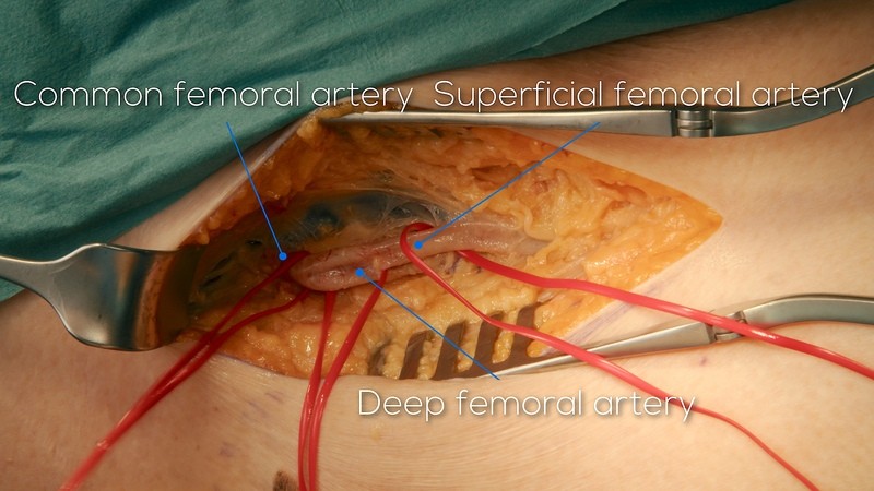
With the vessel cleared of surrounding tissue, the larger caliber of the common femoral artery is visible transitioning to the smaller caliber superficial femoral artery marking the bifurcation point and the deep femoral artery origin.
Controlling the Femoral Arteries
The goal is to gain vascular control proximally at the common femoral artery and distally at the superficial and deep femoral artery level.
A right-angle clamp is used to pass the vessel loop circumferentially around the artery. The instrument passage under the vessel should be done from medial to lateral to avoid injuring the femoral vein wall.
The loop is carefully passed under the artery.
The first vessel to control here is the deep femoral artery, so the loop is passed back and up from the other side of the branch.
Additional exposure of the deep femoral artery can be performed based on the need. To only achieve vascular control of this artery using vessel loops or clamps, this level of exposure is sufficient. However, care should be taken to ensure the vessel loop encircles the main trunk and not merely a side branch. The deep femoral artery gives off branches quite proximally. Distally, the circumflex veins crossing the deep femoral artery are visible. Care must be taken with these as they can cause considerable bleeding if injured.
The superficial femoral artery is looped next.
Then the common femoral artery. The vessel loop is passed twice around the artery, especially on the proximal side, to ensure occlusion in the event of clamp failure or displacement.
The femoral vessels are now controlled with the vessel loops and vascular clamps can now be placed if needed. The patient should be heparinized before vessel occlusion unless active bleeding necessitates immediate vascular control.
Clamp time of the vessels should be minimized. The tolerance to ischemia varies with the clinical scenario and how well the limb is adapted to chronic ischemia. Generally, clamp times up to two hours are considered safe, but the longer the clamp is on, the more it increases metabolic effects during reperfusion.
With vascular control achieved proximally and distally, the vessels are now ready for the intended vascular procedure, whether repair, reconstruction, or endovascular treatment.
Clamp Removal and Closure
Upon completion of the vascular procedure, if vessel suturing was performed, the repair site is vented by allowing blood flow from all directions through the suture line before final tightening of the sutures.
Following this, the vascular clamps are released. Typically, the deep femoral artery clamp is released first, the common femoral artery is then released, creating a preferential pathway for any residual embolic material. The superficial femoral artery clamp is released last. The anesthesia team is informed when clamps are released due to hemodynamic and reperfusion-related metabolic changes.
Drain and hemostatic agent use depends on clinical circumstances. Groin wounds have increased infection risk, especially in peripheral arterial disease patients, which must be weighed against bleeding control needs.
The fascia layer is closed using absorbable suture material with running technique. Care must be taken when placing sutures to ensure accurate bite placement and avoid inadvertent inclusion of femoral nerve branches.
The subcutaneous layer is closed with absorbable continuous sutures. If the great saphenous vein was encountered during dissection, it should not be included in the suture bites during subcutaneous closure.
The skin layer can be closed with interrupted or continuous sutures, or by using surgical staples. Surgical staples, while quick to apply, may pose challenges in cases where there is risk of postoperative bleeding, as they would need to be removed rapidly to allow immediate access to the surgical site for intervention.
To access the full video and additional content on this subject, log in or subscribe
Piftalls and Complications
Pitfalls
Too Distal Incision Site
Using the inguinal crease instead of the true inguinal ligament site as a landmark results in too-distal incision placement, exposing only the superficial femoral artery and missing the bifurcation level.
Incorrect Medial-Lateral Incision Placement
The incision should be centered midway between the ASIS and pubic tubercle. Too medial placement encounters the great saphenous vein, risking femoral vein injury, while too lateral placement encounters the sartorius muscle, risking femoral nerve injury. Encountering these structures indicates the need to adjust the dissection plane. The direction can be adjusted based on the palpable femoral pulse or plaques if present.
Careless Tissue Handling
Inadequate tissue handling in the subcutaneous layer can disrupt lymphatic vessels, leading to persistent lymphatic leakage, seroma formation, and delayed wound healing if these vessels are not clipped during dissection. The superficial veins, including branches of the great saphenous vein system, are also at risk during subcutaneous dissection and should be ligated or preserved.
Rough handling of the femoral vessels during dissection and looping can cause arterial intimal damage, creating tears that predispose to thrombosis.
Aggressive retraction or inadvertent inclusion of the femoral vein during vessel manipulation can result in venous laceration.
Additionally, failure to identify and protect small arterial branches such as the circumflex and epigastric arteries before vessel mobilization can lead to avulsion and bleeding.
Complications
Lymphatic Injury and Leakage
Inadequate control of lymphatic vessels during dissection can result in persistent lymphatic drainage, leading to seroma formation and delayed wound healing. Identification and clipping of lymphatic vessels is recommended, if feasible, as dissection progresses through the subcutaneous layers.
Femoral Vein Injury
The femoral vein lies medial to the artery and can be injured during dissection, retraction, or improper instrument passage. Injury causes massive hemorrhage and is difficult to repair due to the thin vein wall.
Femoral Nerve Injury
Nerve branches lateral to the artery can be injured during dissection or if included in fascial closure sutures, causing motor weakness and sensory deficits.
Circumflex Vein Injury
Veins crossing the deep femoral artery can cause significant bleeding if injured during dissection or vessel mobilization.
Aftercare
General Guidelines
The aftercare protocols may vary between units and depend on type of procedure performed.
Anticoagulation protocols are planned in consultation with vascular surgery. Embolectomy patients typically receive systemic anticoagulation, while arterial repairs may receive antiplatelet therapy.
FAQ
What is the goal of femoral artery exposure?
The goal is to expose and gain vascular control of the common femoral, superficial femoral, and deep femoral arteries for treatment.
Where is the incision positioned?
The skin incision is positioned at the inguinal ligament level, approximately midway between the ASIS and pubic tubercle. The artery lies just medial to the midpoint of the inguinal ligament.
What indicates that the approach has gone too medial?
Encountering the great saphenous vein trunk during dissection indicates the approach has gone too medial and should be redirected laterally.
What indicates that the approach is too lateral?
If muscle fibers become visible beneath the fascia, this indicates the approach is too lateral and dissection should be redirected medially when advancing deeper.
Which vessels are controlled during the procedure?
The common femoral artery, superficial femoral artery, and deep femoral artery are controlled with vessel loops, and vascular clamps can be placed if needed.
In what order are the clamps typically released?
Typically, the deep femoral artery clamp is released first, the common femoral artery is then released, and the superficial femoral artery clamp is released last.
To access the full video and additional content on this subject, log in or subscribe